

 Home
Home


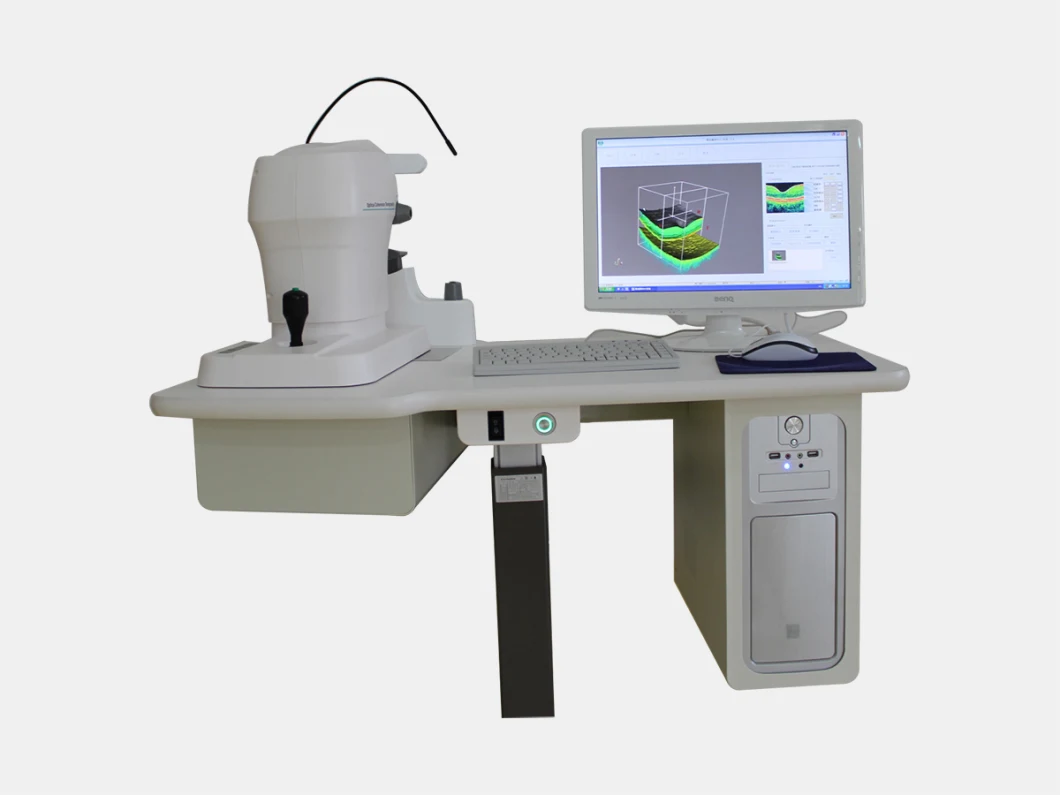
Great Price Ose-2000 Optical Coherence Tomography Oct Machine
Great Price OSE-2000 Optical Coherence Tomography OCT Machine Features: 1. High Performance at a low price: Modular desi
Basic Info
| Model NO. | OSE-2000 OCT Machine |
| Optical Power | ≤ 0.7MW (on The Cornea) |
| Axial Resolution | 5-8 Um in Tissue |
| Lateral Resolution | 15 Um in Tissue |
| Scanners | Galvanometer Mirror |
| Scan Mode | Line, Concentric Ring, Repeat, Arbitrary-Angle |
| Scan Rate | 400 a- Scan/S |
| Acquisition Time | 1 Sec |
| Transport Package | Standard Exporting Packaging |
| Trademark | VSO |
| Origin | Chongqing, China |
| Production Capacity | 1000 Units/Month |
Product Description
Great Price OSE-2000 Optical Coherence Tomography OCT MachineFeatures:
1. High Performance at a low price: Modular design increase flexibility, reusability and maintainability. We can provide personalized design according to the customer's needs.
2. With the powerful software, OSE-2000 has clear, easy to use interface and support multi-language.
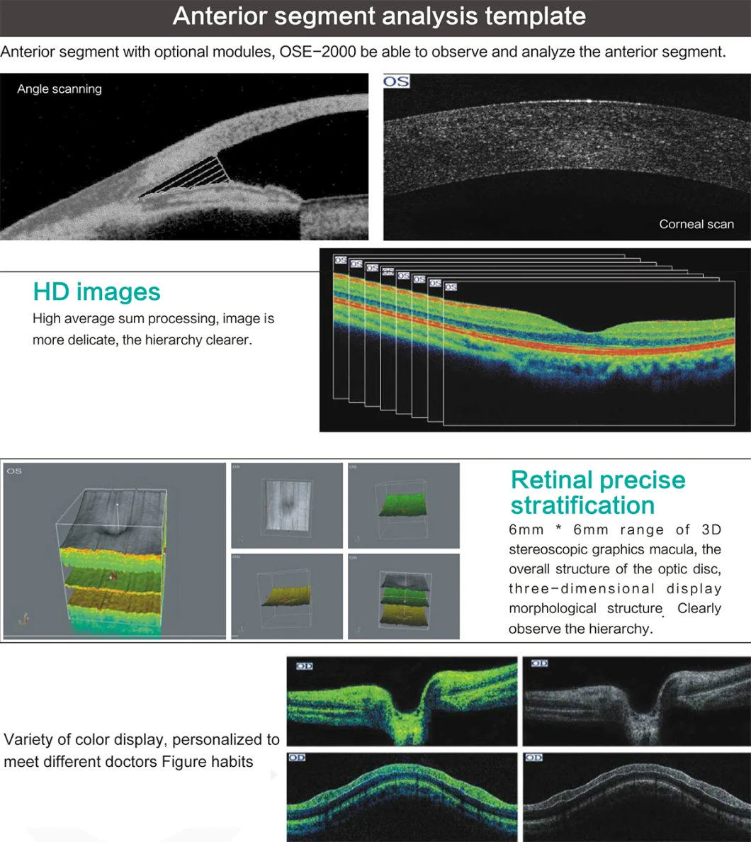
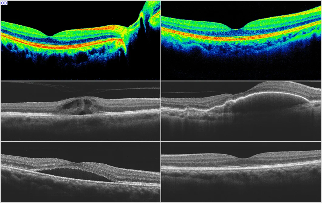
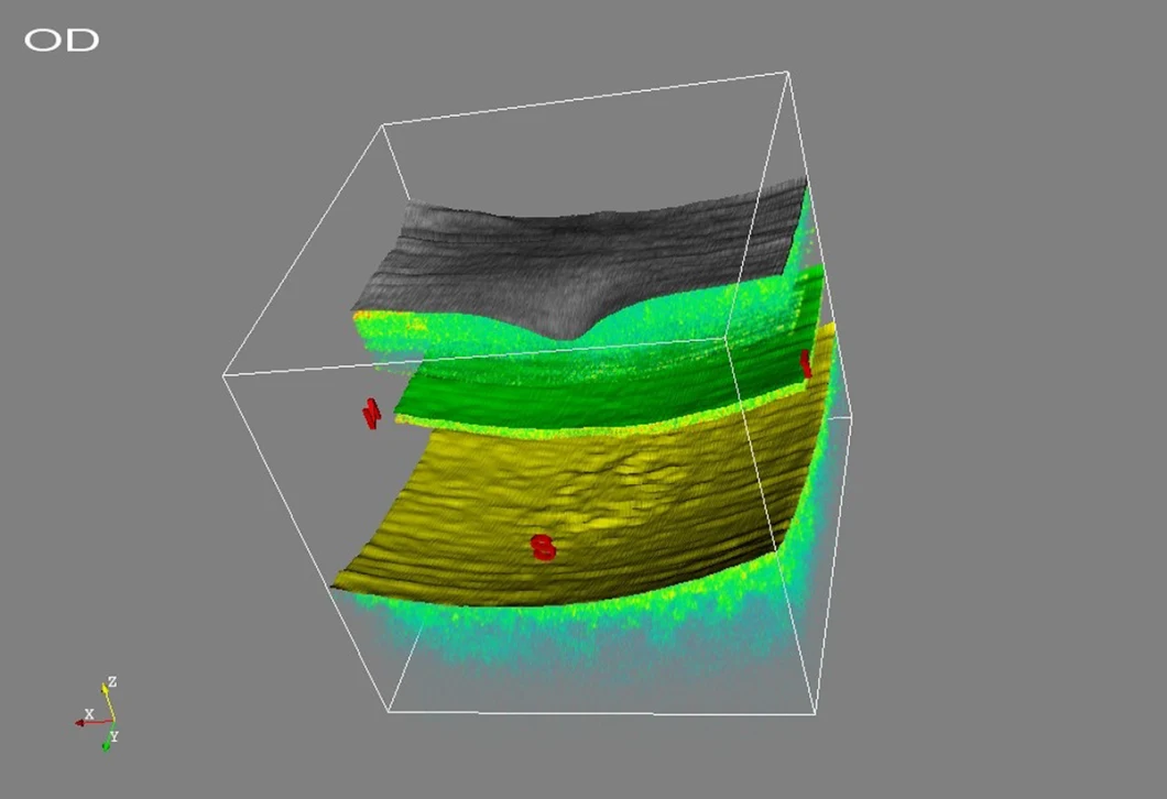
3. Anterior segment analysis template: anterior segment with optional modules, OSE-2000 be able to observe and analyzer the anterior segment.
Specifications:
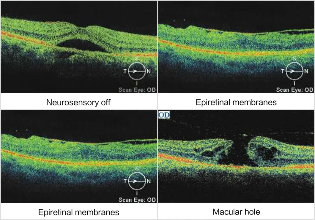
| Tomographic Imaging | |
| Purpose | Cross sectional imaging of the retina |
| Signal type | Photon scattering from tissue |
| Light source | Super luminescent diode, 830nm |
| Optical power | ≤ 0.7mW (on the cornea) |
| Axial resolution | 5-8 um in tissue |
| Lateral resolution | 15 um in tissue |
| Scanners | Galvanometer mirror |
| Scan mode | Line, concentric ring, repeat, arbitrary-angle |
| Scan rate | 400 A- scan/s |
| Acquisition time | 1 sec |
| Scan depth | 2mm in tissue |
| Fundus Imaging | |
| Purpose | Fundus observation and real-time registration of OCT imaging |
| Signal type | CCD imaging |
| Field angle | 29°x 23° |
| Viewing method | 15-inch color flat panel display |
| Illumination | LED |
| Internal fixation | LED dot matrix |
| External fixtion | Adjustable blinking LED |
| Minimum pupil diameter | 3.5mm |
Send to us